Last year, only 21 new drugs were approved for use by the US Food and Drug Administration (FDA), down from 26 in 2009 and a huge drop from the late-1990s when as many as 53 new molecular entities were given the go ahead in a single year. The decline is so noteworthy that the US federal government has announced plans to stimulate the creation of new treatments by launching its own drug development centre, the National Center for Advancing Translational Sciences (NCATS). This initiative comes at a time when many of the world’s leading pharmaceutical companies have been shrinking their research budgets. The New York Times recently reported that this slowdown in funding means that even some promising new discoveries are not being pursued — a situation that could affect millions of people, suffering from cancer, HIV/AIDS and thousands of other illnesses, whose lives depend upon new scientific advances.
Reducing the high cost of drug testing methods – while also improving their accuracy – could help reverse this trend. Safety and efficacy testing for a single drug compound using traditional animal models can take years to complete and often costs more than $2m. Moreover, these controversial testing methods often do not reliably predict human responses. Forexample, in describing the proposed NCATS, Francis S. Collins, director of the US National Institutes of Health (NIH), told attendees at the Clinical Research Foundation annual meeting in April 2011 that the ‘low concordance between animal and human toxicity’ is a constriction point in preclinical toxicity testing. For this reason, many new compounds pass preclinical trials in animal models, only to end up failing miserably when they reach clinical trials in humans.
A more reliable, timely, and economical testing method could dramatically accelerate the arrival of affordable and promising new drug discoveries on the market. This would also have great value for safety testing of environmental toxins, chemical and nanoparticulates. In fact, scientists have been working for many years to create such alternatives to animal models, but with only moderate success.
Proof-of-concept
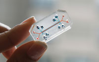
At the Wyss Institute for Biologically Inspired Engineering at Harvard University, we recently developed a technology that is poised to address this critical need. We have found a way to replicate complex organ structures by using patterns microetched into flexible biocompatible materials. We have used microfabrication techniques that were initially developed to manufacture microchips for the computer industry. The patterns can be shaped into microfluidic systems containing hollow channels lined by living human cells that are fed by flowing culture medium. As proof-of-concept for this ‘organ-on-a-chip’, we microengineered a device that replicates the three-dimensional architecture of the human lung, as well as its physical movements.
As described in a recent issue of Science (2010, 332 , (6038), 1662), this microdevice is a clear flexible chip about the size of a computer memory stick and contains hollow microchannels lined by living human lung cells. The device simulates many of the major functions of the human lung air sac that are critical for health, including gas exchange, chemical absorption, and immune responses to infection. These capabilities make the device ideally suited to testing drug safety and efficacy, as well as assessing the absorption and toxic effects of airborne nanoparticles.
This living lung-on-a-chip consists of three parallel microfluidic channels etched into a rubbery, transparent polymer composed of polydimethylsiloxane. This configuration mimics the essential structure of the alveoli, the air-filled microscopic cavities deep inside the lung surrounded by capillary blood vessels where the exchange of oxygen and carbon dioxide takes place. The central channel in the microdevice is divided horizontally by a clear porous flexible membrane coated with extracellular matrix molecules similar to those that hold together the tissues of the air sac and the adjacent blood capillaries in the living lung. In the device, human lung lining cells are cultured on the top of the membrane and air is flowed above them, while human capillary blood vessel cells from the lung are cultured on the opposite side of the same membrane. Culture medium is flowed through the space beneath the capillary cell layer to mimic blood flow through lung microvessels.
In addition to replicating the structure of the most critical portion of the human lung, this biomimetic microsystem copies its mechanical behaviour as well, in that it can ‘breathe’. This breathing takes place as the air pressure in the two side channels is alternatively decreased and increased by using a small vacuum pump. This action causes the human cell layers to stretch as the central membrane widens and then contract as the membrane contracts. These movements precisely mimic the motion experienced by cells in the air sac of a human lung.
To determine how well the device replicates the natural responses of living lungs to physiological stimuli, we infected the air space in the device with living bacteria, while flowing white blood cells through the channel on the blood vessel side. The lung cells detected the bacteria and, through the porous membrane, activated the underlying blood vessel cells, which in turn triggered an immune response. This caused the white blood cells to adhere to the vessel cells, squeeze through the pores in the flexible membrane and move into the air chamber where they engulfed the bacteria.
We also explored the potential use of this microdevice for environmental toxin testing. Silica nanoparticles on the scale of a few billionths of a metre, similar to common air pollutants, were introduced into the air sac channel. These nanoparticles entered the lung cells and caused the cells to overproduce free radicals and to induce inflammation. The response was highly sensitive to physiological breathing movements, a result that would never have been detected with conventional tissue culture models.
Many of the particles were transported across the lung tissue layers and passed into the blood channel, and again, mechanical breathing greatly enhanced the absorption of these nanoparticles.
Impressively, this novel finding that mechanical distortion of living human tissues increases absorption of nanoscale toxicants was confirmed in whole lung studies in mice. This is an important finding because manmade nanoparticulates now appear in air and water pollution, as well as in cosmetics, electronics, and household cleaners, yet their long-term health effects are not well understood.
In the case of lung-on-a-chip, the goal is to develop in vitro models for key lung diseases, such as pulmonary oedema, injury, fibrosis, asthma, chronic obstructive pulmonary disease, and cystic fibrosis, where current animal models fail to predict human responses or are extremely complex, costly and time-consuming. The data generated will allow the Institute to measure the value of this approach using drugs currently in the clinic and compounds under development provided by the pharmaceutical industry as benchmarks of success. Compounds that were found to be safe in animals, but failed in human trials, will be of particular value for these validation studies. The Institute is also pursuing discussions with companies in other sectors, including consumer healthcare products and chemical products, as well as with start-ups.
A heart–lung micromachine
At the Wyss Institute, which operates as an alliance between Harvard’s schools and affiliated hospitals, Boston University and the University of Massachusetts Medical School, we are now applying the organ-on-a-chip technology to develop a heart–lung micromachine with $3.3m funding from the FDA and NIH. This device is designed to replicate the coupled complex physiological functions and mechanical microenvironments of a breathing lung and beating heart. This should provide accurate and immediate measures of the efficacy and safety of inhaled drugs, nanotherapeutics and other medical products on integrated lung and heart function.
The FDA/NIH-sponsored heart–lung micromachine project represents a paradigm shift in that the FDA is working with the Wyss Institute more as an active collaborator than as a grant management organisation. This ability to work closely with the FDA early into the device development process should increase the likelihood of technical success, while also enhancing the acceptance of these new organ-on-a-chip technologies.
Future prospects
The Wyss Institute is working on other organ models that could have high-impact applications as well. A more predictive human kidney-on-a-chip, for instance, would be a valuable tool for investigating absorption, distribution, metabolism, excretion and toxicological properties of new chemical entities during drug development. We are also working on an intestine-on-a-chip that exhibits peristalsis: the series of muscle contractions that occur as food moves through the digestive tract. The intestine is the major site for absorption of orally-delivered drugs, foods and nutriceuticals. Intestinal epithelial cells have drug metabolising enzymes and drug transporters that play a critical role in determining distribution, metabolism, excretion and toxicity of various chemical compounds that influence bioavailability and potential drug–drug interactions. Bone marrow and cancer models are being developed for additional screening applications and an artificial spleen-on-achip is also being explored as a sepsis therapeutic device and a rapid blood infection diagnostic.
The market need is so great for this technology that the Wyss Institute is working actively to translate it into products and therapies with immediate applications through corporate alliances, and potentially through new start-ups. The Institute is initiating collaborations with several pharmaceutical companies to help demonstrate its use and validate the model for key drug-development applications.
The potential for this approach is huge and investigators around the world are beginning to explore similar organ-on-a-chip alternatives to animal testing. One can imagine someday linking many different human organ mimics together, such as artificial microscale lungs, hearts, livers, kidneys, intestines, skins and bone marrows, through microengineered vascular channels. This ‘human-on-a- chip’ could allow one to measure absorption and bioavailability of drugs delivered by oral, aerosol, transdermal or intravenous routes, while analysing clearance by the liver and kidney and screening for toxicity and immune reactions across multiple organ systems. It might be possible to use these integrated biochips to carry out ‘human clinical trials on chips’ in which human cells isolated from genetically distinct human populations with known sensitivity to particular types of chemical agents could be selected for, or excluded from, testing of high value agents similar to human trials in the clinic. It is unlikely that this approach will ever fully replace animal or human testing, but the possibility of shortening the time and cost for drug development, and for increasing the likelihood for success in human trials, offers great potential for transformative change in the pharmaceutical and chemical industries.
Donald E. Ingber is founding director of the Wyss Institute for Biologically Inspired Engineering at Harvard University, US.


