Each year SCI’s Scotland group a runs competition where students are invited to write a short article describing how their PhD research relates to SCI’s strapline: where science meets business.
Anastasia Kapara (right), a Chemistry PhD student at the University of Strathclyde, is the first of this year’s four winners. Her article ‘Distinguishing breast cancer cells using innovative medical imaging approaches’ is reproduced here:
Distinguishing breast cancer cells using innovative medical imaging approaches
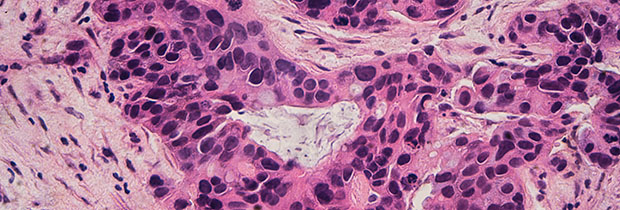
Microscopic image of Grade 2 breast cancer cells.
Every two minutes a woman is diagnosed with breast cancer, which makes the disease one of the leading cause of death among women. Clinical outcomes have shown that patients with positive Estrogen Receptor alpha (ERα+) or Human Epidermal Growth Factor Receptor 2 (HER2+) breast cancer are generally associated with a poorer prognosis, drug resistance, and a higher rate of disease recurrence. Therefore, it is crucial to detect and identify these two biomarkers in breast cancer cell populations for therapeutic decision making and determination of the clinical outcome.
Although there are many publications that try to mitigate the limitations of breast cancer identification, their methods remain on the paper. Thus, the main question here is ‘What is the problem with these research approaches’? Although the answer can not be simple, it has been shown that a well constructed and multi-disciplinary plan, that engages science and business, is missing. Therefore, it is vital for current publications to have a major impact on the society rather than just the scientific world.
My PhD research tries to move from a clear scientific background to an entrepreneurial healthcare approach. Specifically, in my project I use surface-enhanced Raman spectroscopy (SERS) for examining the breast cancer disease. SERS offers higher specificity, selectivity and simultaneous screening of molecules for the cell phenotype characterisation, in comparison to conventional imaging techniques. Here, the main aim is to create an innovative imaging platform for targeting, detecting and tracking ERα and HER2 in breast cancer cells simultaneously, using SERS combined with functionalised gold nanoparticles (AuNPs). Most importantly, the project offers strong ties with industry as it suggests that a simple and efficient method can be used to identify and track the cancer cells. Therefore, this research proposes to deliver cutting edge technologies and modern therapeutics with tendencies to commercialisation.

For the evaluation of this scientific idea and for a translational research (‘bench to bedside’ approach), my project is funded by OPTIMA CDT, hosted by the University of Edinburgh and University of Strathclyde. This programme places emphasis on interdisciplinary opportunities into innovative healthcare technologies and advanced business analytics applications, which has helped me to enrich my transferable skills and my current research outside of the academic lab. This practical exposure to healthcare entrepreneurship has introduced me to principles that no book can teach.
Through my PhD project, I have taken classes in entrepreneurial finance, innovation-driven entrepreneurship, translational studies, ethics and regulation, as well as technology commercialisation and entrepreneurial ecosystem. The marriage of business development and science has helped me to understand the investigation of the decision-making process in a project and the evaluation of its innovative aim.
Currently, the experimental results indicate that the functionalised gold nanoparticles have low cellular toxicity and good biocompatibility in breast cancer cells. The SERS cell mapping revealed that there is a strong targeting effect of the probes into different cell lines based on their functionalisation properties. Parallel experiments showed that there is lower probe accumulation in cells treated with drugs that induce degradation. These data show that the novel SERS probes could be utilised to distinguish cancer cells for direct molecular imaging and quantification of ERα and HER2 in living breast cancer cells, and inform about the efficiency of drug candidates, with the advantages of high SERS sensitivity, and biocompatibility. As a result, there are major opportunities here for the creation of innovative platforms that combine science and business for a modern therapeutic approach.