Careers
SCI’s America International group has awarded the 2021 Perkin Medal to Dr Jane Frommer. The 114th Perkin Medal was presented to Jane at the Bellevue Hotel in Philadelphia, Pennsylvania, in recognition of her outstanding contribution to chemistry.
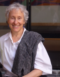
Dr Jane Frommer
Dr Frommer is renowned for her key contributions in electronically conducting polymers and scanning probe instrumentation. Her pioneering work with scanning probes paved the way for their use in chemistry, materials science and, eventually, in nanotechnology. According to SCI America, her nanoscopic analytic methods are vital to nanostructural research and are used across many industries.
Dr Frommer began her career in 1980 at Allied Corporate Laboratories (now Honeywell), where she created the solution state of electronically conducting organic polymers. In 1986, she joined IBM where, along with other instrumentalists, she demonstrated the ability to image and manipulate single molecules using scanning tunnelling microscopy. During her multi-year assignment at the University of Basel Physics Institute in the early 1990s, Dr Frommer’s team expanded the capability of scanning probes in measuring the functional properties of organic thin films with atomic force microscopy.
Since 2018, she has worked as a science advisor for Google. In this capacity, she has sought to increase the amount of open source data available in the physical and life sciences. She also helps Silicon Valley start-ups navigate the chemical and material challenges of nanotechnology and has mentored countless students and young scientists in high school, college, and in her laboratory in recent decades.
Previous recipients of the Perkin medal include Barbara Haviland Minor, of the Chemours Company, and Ann E Weber, of Kallyope Inc.
Dr Frommer has written more than 100 referred publications and is the co-inventor of more than 50 issued patents. With her extraordinary body of work spanning more than 40 years, she is a worthy recipient of the prestigious Perkin Medal.
The Perkin Medal is widely acknowledged as the highest honour in American industrial chemistry. It was established to commemorate the 50th anniversary of William Henry Perkin’s discovery of mauveine at the age of just 18. Perkin’s creation of mauveine, the world’s first synthetic aniline dye, revolutionised chemistry and opened up new frontiers in textiles, clothing, and other industries. Perkin was a founding member of SCI and this Medal was first presented to him in New York in 1906.
For more information on the Perkin Medal and the nomination process, visit: soci.org/awards/medals/perkin-medal
30 September 2021 |
|


 |
|

Science & Innovation
Every day, there are subtle signs that machine learning is making our lives easier. It could be as simple as a Netflix series recommendation or your phone camera automatically adjusting to the light – or it could be something even more profound. In the case of two recent machine-learning developments, these advances could make a tangible difference to both microscopy, cancer treatment, and our health.
The first is an artificial intelligence (AI) tool that improves the information gleaned from microscopic images. Researchers at the University of Gothenburg have used this deep machine learning to enhance the accuracy and speed of analysis.
The tool uses deep learning to extract as much information as possible from data-packed images. The neural networks retrieve exactly what a scientist wants by looking through a huge trove of images (known as training data). These networks can process tens of thousands of images an hour whereas some manual methods deliver about a hundred a month.
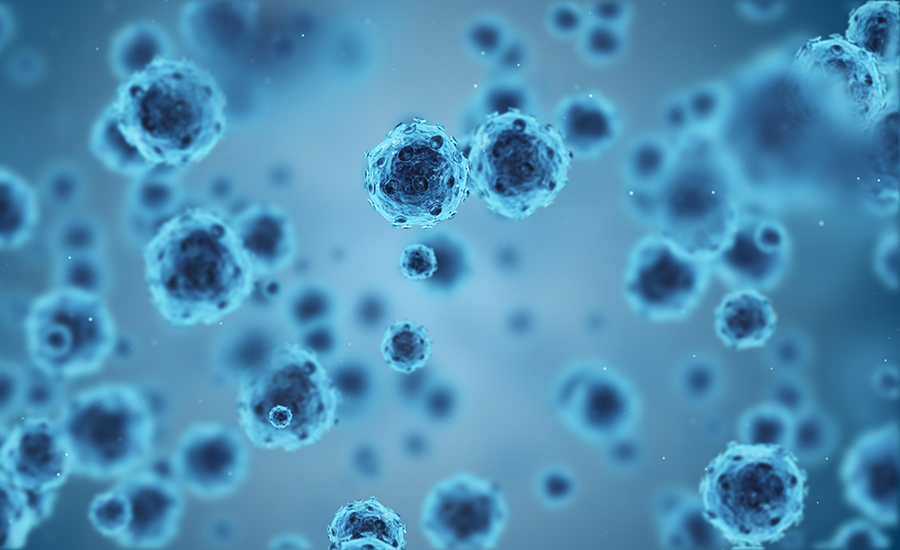
Machine learning can be used to follow infections in a cell.
In practice, this algorithm makes it easier for researchers to count and classify cells and focus on specific material characteristics. For example, it can be used by companies to reduce emissions by showing workers in real time whether unwanted particles have been filtered out.
“This makes it possible to quickly extract more details from microscope images without needing to create a complicated analysis with traditional methods,” says Benjamin Midtvedt, a doctoral student in physics and the main author of the study. “In addition, the results are reproducible, and customised. Specific information can be retrieved for a specific purpose."
The University of Gothenburg tool could also be used in health care applications. The researchers believe it could be used to follow infections in a cell and map cellular defense mechanisms to aid the development of new medicines and treatments.
Machine learning by colour
On a similar thread, machine learning has been used to detect cancer by researchers from the National University of Singapore. The researchers have used a special dye to colour cells by pH and a machine learning algorithm to detect the changes in colour caused by cancer.
The researchers explain in their APL Bioengineering study that the pH (acidity level) of a cancerous cell is not the same as that of a healthy cell. So, you can tell if a cell is cancerous if you know its pH.
With this in mind, the researchers have treated cells with a pH-sensitive dye called bromothymol blue that changes colour depending on how acidic the solution is. Once dyed, each cell exudes its unique red, green, and blue fingerprint.
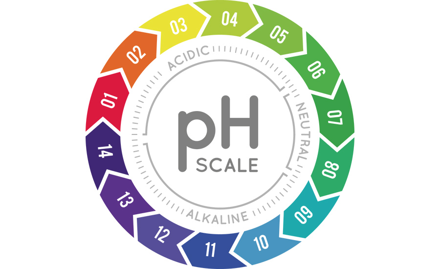
By isolating a cell’s pH, researchers can detect the presence of cancer.
The authors have also trained a machine learning algorithm to map combinations of colours to assess the state of cells and detect any worrying shifts. Once a sample of the cells is taken, medical professionals can use this non-invasive method to get a clearer picture of what is going on inside the body. And all they need to do all of this is an inverted microscope and a colour camera.
“Our method allowed us to classify single cells of various human tissues, both normal and cancerous, by focusing solely on the inherent acidity levels that each cell type tends to exhibit, and using simple and inexpensive equipment,” said Chwee Teck Lim, one of the study’s authors.
“One potential application of this technique would be in liquid biopsy, where tumour cells that escaped from the primary tumour can be isolated in a minimally invasive fashion from bodily fluids.”
The encouraging sign for all of us is that these two technologies are but two dots on a broad canvas, and machine learning will enhance analysis. There are certainly troubling elements to machine learning but anything that helps hinder disease is to be welcomed.
Machine Learning-Based Approach to pH Imaging and Classification of Single Cancer Cells:
https://aip.scitation.org/doi/10.1063/5.0031615
Quantitative Digital Microscopy with Deep Learning:
https://aip.scitation.org/doi/10.1063/5.0034891
23 March 2021 |
|
|