The rise of the lab-on-a-chip offers a host of benefits to analytical scientists. By making analytical instruments smaller, faster, more sensitive and able to work with much smaller quantities of chemicals, it allows the scientists to get out of the laboratory and conduct on-the-spot analyses in real time. But these benefits come at the expense of a whole load of challenges, particularly for lab-on-a-chip versions of capillary electrophoresis (CE).
A mainstay of chemical analysis for over 40 years, CE provides a way to separate charged particles or molecules based on their size and charge. It involves fi lling a long capillary with an electrolyte and then placing electrodes at either end before passing a current through it. Any charged particles or molecules placed in the capillary are then pulled through the electrolyte towards the electrode with the opposite charge.
If the particles differ in ionic charge, then they can be separated in a liquid electrolyte, because those with a stronger charge will migrate faster than those with a weaker charge. If the particles possess charges with similar strengths but differ in size then the electrolyte needs to be a thick gel, which will impede larger particles more than smaller ones.
This kind of gel-based CE has proved particularly adept at separating biomolecules, such as DNA, which are naturally negativelycharged, and proteins, that can be negatively- or positively-charged depending on the pH of the surrounding solution. As such, gel-based CE is commonly employed in gene sequencing and proteomics studies.
Although CE has been shrunk to fit onto a single chip, with commercial versions already being sold by companies such as Agilent Technologies and Caliper Life Sciences, such microchip CE systems are not without their problems. Conventional CE utilises capillaries that are a few tens to hundreds of micrometres wide and around 50cm long. The separation channels in microchip CE are a similar width but are only a few centimetres long. Because of the much shorter length, microchip CE systems have struggled to match the separation effi ciency of conventional CE.
On top of this, actually inserting thick gels into tiny channels can be quite a challenge, like trying to push jelly into a straw. These problems are encouraging researchers to come up with alternatives to CE that are more suitable to operating at small scales, some of which could soon fi nd themselves in the hands of analytical scientists.
Perhaps the most obvious option is simply to replace the gel with a material that is both more effective at separating biomolecules at small scales and easier to insert into tiny channels, but fi nding such a material has proved far from easy. Then, last year, a team of chemists from Kent State University in Ohio, US, led by Oleg Lavrentovich, showed that liquid crystals might be just such a material.
As their name suggests, liquid crystals are liquids made up of molecules that display some form of crystal-like order. Being liquids, they can easily be inserted into tiny channels, while their crystal-like order ensures that particles interact with them in some very interesting ways.
This, at least, is what Lavrentovich and his team discovered when they tried conducting electrophoresis in a tiny channel fi lled with a liquid crystal consisting of lots of long, rod-like molecules lined up horizontally. When they applied a current, they found that both charged silica particles and uncharged gold particles would migrate through the channel. Normally, you would only expect the charged particles to move.
But that’s not all, because the particles moved in the same way irrespective of whether Lavrentovich applied a direct or an alternating current. Normally, electrophoresis can only be conducted using a direct current, because an alternating current simply causes the charged particles to jiggle about rather than move in a single direction.
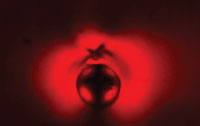
Investigating this effect, Lavrentovich found that it was all down to the way the particles disturb the orientation of the rod-like molecules. ‘When a sphere is placed in a liquid crystal, it distorts the molecular orientation,’ explains Lavrentovich. ‘It's like the sphere acquires a little “tail” or a “fin”. This tail helps to propel the particle in the liquid crystal.’ Lavrentovich is now exploring this effect further and is also looking at using it to separate mixtures of particles.
An alternative way to replace gel electrolytes with liquid electrolytes is to fi ll the channel with some kind of physical structure. For example, a number of research groups are fi lling the channels with arrays of microscopic pillars.
These micropillars can separate particles in two distinct ways. They can physically impede the particles as they make their way along the channel, with large particles impeded more than small particles, or the particles can take different trajectories between the micropillars, with the precise trajectory again dependent on size. Such micropillars have proved adept at separating a wide range of different particles, including DNA, proteins, inorganic molecules and even whole cells.

As liquid gradually evaporates from the open end, the nanoparticles weld together to form a solid wall that fi lls the channel. But this wall is permeated by small gaps between the spherical nanoparticle ‘bricks’, producing a network of pores that provide a tortuous pathway through the wall. Furthermore, the size of those pores can be controlled by simply varying the size of the nanoparticle bricks: larger nanoparticles produce larger pores.
The ease with which particles can pass through this wall obviously depends on their size, allowing them to be separated. Harrison has shown that such walls can effi ciently separate a wide range of different size proteins and DNA strands.
For some particles, you don’t even need to go to the trouble of building pillars or walls; a simple fl at surface will do just as well. Miriam Rafailovich and her colleagues at the State University of New York (SUNY) at Stony Brook , US, have discovered that different length DNA strands move at different speeds across a flat silicon surface covered in a liquid electrolyte.
This is because friction causes the DNA strands to stick to the surface as they move across it, with larger strands sticking more than shorter strands. This will happen on a bare silicon surface, but Rafailovich found the effect could be enhanced by covering the bare surface with gold dots around 200nm in diameter.
Rather than finding novel replacements for electrophoresis gels, some researchers are investigating novel forms of electrophoresis, such as dielectrophoresis and acoustophoresis.
In CE, passing a direct current through the capillary sets up a uniform electric field that induces movement in charged particles. In contrast, dielectrophoresis utilises a non-uniform electric field, which varies in strength along the channel, to induce movement in both charged and uncharged particles. This non-uniform field can be generated by arrays of electrodes spread along the channel or small areas of non-uniform field can be generated by placing insulating structures in a uniform field.
Placing a particle in an electric field causes the negative electrons and positive atomic nuclei in the particle to be pulled towards opposite ends, producing what is known as a dipole moment. In a uniform field, this has no effect on the particle, but in a non-uniform electric field it causes the particle to move either towards or away from the strong region of the electric field. Exactly how the particle moves depends on the magnitude of its dipole moment, which is determined by the interaction between numerous factors, including its size, shape, charge and polarisability.
A number of research groups have found ways to use dielectrophoresis to separate particles, but this separation process works differently to CE. Rather than particles moving at different rates along a capillary, dielectrophoresis separates particles by trapping them at specific positions along a channel or guiding them into different channels.
Some researchers have managed to combine electrophoresis and dielectrophoresis on a single microchip, giving them extremely fine control over the movement of different size particles. Mark Hayes and his colleagues at Arizona State University in Tempe, US, recently did this by filling a capillary with two rows of insulating spikes shaped like sharp teeth that don’t quite meet in the middle.
Applying a current along the channel sets up a uniform electric field that causes both charged and uncharged particles to move along the channel; the uncharged particles are carried along by electro-osmosis, which is a steady flow in the liquid electrolyte generated by the field. When they reach the spiky region of the capillary, however, the uniform field becomes non-uniform, allowing dielectrophoresis to take over and trap the particles in the spaces between the spikes.
Exactly when this happens depends on the magnitude of the particles’ respective dipole moments and the size of the gaps between the teeth, which gradually decrease along the channel. The upshot of this is that when Hayes and his team sent different size polystyrene particles down the channel, they separated according to size, with different size particles becoming trapped between spikes at different places along the channel. In general, larger particles ended up further along than smaller particles.
Rafailovich, meanwhile, is now working on ways to combine dielectrophoresis with her sticky silicon surfaces. ‘The goal is to create an integrated chip where broad bands of DNA are separated into sub-regions where an external field can be turned on for finer separations,’ she says.
Other researchers are looking to replace the electric current with sound waves. The idea is to project ultrasound at particular frequencies into a channel etched into a microchip, creating stable areas of high and low pressure. Particles in the channel are then attracted towards either the high or low pressure areas.
In general, hard, rigid particles move towards the low pressure areas and softer, more compressible particles move towards the high pressure areas. By tailoring the ultrasound frequency, researchers can induce a low pressure region right along the middle of the channel and high pressure regions along the two side walls. When a mixture of particles is pumped through this channel, the rigid particles congregate in the middle of the channel while the soft particles congregate along the outer walls, separating them from each other.
Thomas Laurell at Lund University in Sweden is one of the pioneers of this approach and has successfully used acoustophoresis to remove soft fat molecules from both blood and milk.
Although many of these novel forms of electrophoresis have yet to leave the laboratory, some are just starting to find their way into commercial lab-on-a-chip systems. For example, Swedish company ErySave is looking to commercialise Laurell’s acoustophoresis microchip, which it envisages being used to clean blood during open-heart surgery.
Meanwhile, the Belgian nanotechnology company imec is utilising dielectrophoresis and micropillars in two lab-on-a-chip devices it is developing as part of its Human++ programme.
Imec is using dielectrophoresis to help separate cancer cells from blood cells in a lab-on-a-chip device for monitoring cancer progression and the effect of treatment. While, in conjunction with the Japanese electronics giant Panasonic and the Vrije Universiteit Brussel, it is using an array of micropillars to separate DNA strands in a lab-on-a-chip device for detecting specific single nucleotide polymorphisms (SNPs), which are places on the human genome that vary between people. These micropillars are also coated with an ion-exchange material, which means they actively interact with the DNA strands rather than simply impeding their progress.
‘You get much better separation of the various DNA strands with these ordered micropillars,’ explains Maaike Op de Beeck, Human++ program manager at imec. ‘Otherwise you’d need much longer filters to get the same separation and much more fluid.’
It’s CE, but not as we know it.
Jon Evans is a freelance science writer based in Chichester, UK.


