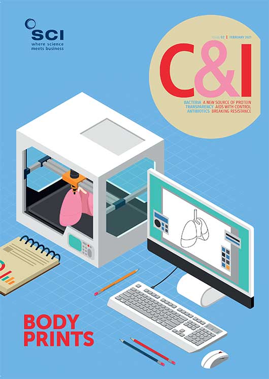
Bioprinting is an emerging technology that produces 3D anatomical-scale structures that imitate natural tissues, Maria Burke reports
With their pre-programmed structures and geometries, bioprinted tissues have various applications for replacing injured or diseased tissues. As well as tissue engineering, they also show promise for drug discovery, toxicology assays and in vitro models of disease.
The ‘bioinks’ used to make tissue constructs are solutions of a biomaterial, or a mixture of several biomaterials, encapsulating the desired cell types. They may be made from natural or synthetic biomaterials, or a combination of the two. The development of new bioink materials and the engineering of novel bioink formulations are currently major areas of interest.
To create tissue scaffolds from bioinks, cells and growth factors are added to liquid biomaterials, which solidify on deposition. The cells start to produce new proteins around the scaffold, eventually creating structures that resemble natural tissues, such as a patch for a lung, or an artificial material such as a dialysis membrane.
Bioinks have to be biocompatible and, if necessary, biodegradable. They need to provide a cell-friendly environment, and produce constructs that are strong and robust, and keep their shape. Various biomaterials make good bioinks, usually in the form of hydrogels. They offer several advantages for scaffold materials as they are biocompatible and typically biodegradable, and many have specific cell-binding sites useful for cell attachment, growth and differentiation. Hydrogel biomaterials include protein-based ones such as gelatin, collagen, fibrinogen and silk; polysaccharides such as alginate, gellan gum, chitosan and hyaluronic acid; and synthetic materials such as polyethylene glycol.
Biomedical engineers at Rutgers University-New Brunswick, US, have been studying hyaluronic acid (HA), a natural molecule – a non-sulfated glycosaminoglycan – found in many tissues. It plays a significant role in synovial fluid, vitreous humour and cartilage. When dissolved in water, the HA solution becomes viscous, making it a promising material for applications as bone and cartilage constructs. But, while it has the right properties for scaffold-building, it lacks durability. The team’s solution was to combine thiol-modified hyaluronic acid with polyethylene glycol diacrylate. The combined substances form a strengthened gel.1
With this approach, the team believes it can print tougher gel scaffolds with varying levels of stiffness. Like traditional printers that rely on four pigments to cover the whole colour spectrum, their system would include hyaluronic acid and polyethylene glycol as the basic ‘ink cartridges’ and add in other cartridges, featuring inks with different cells and ligands that serve as binding sites for cells.
‘Both the stiffness and the binding sites provide important signals to cells,’ explains lead author Madison Godesky. ‘What especially distinguishes our work from previous studies is the potential to control the stiffness and ligands independently through combinations of inks.’
US researchers have created two specialised composite hydrogel bioinks used to print a type of cartilage. Known as fibrocartilage, it helps connect tendons, ligaments or bones. The best formula was made into structures implanted safely into mice for 10 weeks.
When bioprinted algae and human liver-derived cells were embedded in a 3D hydrogel, the algae released oxygen during photosynthesis, and enhanced the viability and functions of the human cells, which grew well and produced liver-specific proteins.
Akhilesh Gaharwar’s team at Texas A&M University, US, is also working on developing more durable structures. They have designed a new class of hydrogel also made from polyethylene glycol but this time combined with nanoparticles. Their Nanoengineered Ionic-Covalent Entanglement (NICE) hydrogels benefit from two reinforcement mechanisms that appear to work together to improve mechanical strength, elasticity, toughness and flow properties. The team also notes that ‘a unique aspect’ of the NICE bioink is its ability to print much taller structures than can be achieved with conventional bioinks without requiring secondary supports.
Gaharwar’s team reported recently that their cell-laden NICE bioinks deposit new proteins, rich in a cartilage-like matrix, which then calcifies to form mineralised bone over a three-month period.2 Almost 5% of the printed scaffolds consisted of calcium, which is similar to cancellous bone, the network of spongy tissue typically found in vertebral bones. Such scaffolds show promise as patient-specific bone grafts offering new treatments for patients suffering from arthritis, bone fractures, dental infections and craniofacial defects.
Spider silk
Another attractive bioink is spider silk because it is relatively safe to use in the body and is extraordinarily strong. New research confirms its promise as 3D printed hydrogel scaffolds for tissue regeneration because it both repels microbes and encourages human cells to grow and regenerate. Remarkably, most spider silk webs remain resistant to microbial decomposition for years. But how they manage this remains unclear.
Researchers from the University of Bayreuth in Germany have been studying this microbe-resistance in recombinant spider silk proteins. Compared with natural spider silk, they found that only the structural features of individual recombinant proteins are necessary to generate a microbe-repellent surface; no additional components, such as glycoproteins, lipids or antimicrobial agents, are involved.3
‘The decisive factor lies in structures at the nanometre level,’ says team leader Thomas Scheibel. ‘To the best of our knowledge, this is a completely new finding, which opens the door for novel applications of spider silk materials, for example, for the preparation of bioinks for biofabrication and regenerative medicine.’
Working with a composite of proteins and graphene oxide, researchers have combined 3D printing with self-assembly to create tubular structures that replicate some properties of vascular tissue.
Silk is also proving useful to help various bioinks retain structural integrity and 3D configurations after printing. Considerable research efforts are focused on how to fix cells onto the scaffold after printing. There is a risk that cells get damaged during printing or produce structures that don’t maintain the desired shape.
Researchers from Osaka University in Japan added silk nanofibres to different bioinks. In one experiment, they bioprinted a nose-like configuration that retained its shape only when printed with bioink containing the silk fibres.4 The team speculates the silk fibres minimise high mechanical stresses on cells during printing. For example, Young’s modulus – a measure of stiffness – increased several-fold and remained enhanced for over a month.
To produce the silk fibres, the team first removed sericin from virgin silk – because this protein causes inflammation in patients – and then ground the remaining biocompatible material into nanofibres, which are then sterilised. They report that over 85% of the cells in the bioink remained alive after a week in the printed bioink with or without the added fibres, indicating that the fibres did not damage the cells.
‘Our silk fibres are excellent additives to bioink cell printing media,’ says lead author Shinji Sakai. ‘They are compatible with many media, such as those containing gelatin, chitosan, or hyaluronic acid, giving them a broad range of potential applications.’
Composites
Composite bioinks are another hot topic. For example, US researchers at the Wake Forest Institute for Regenerative Medicine (WFIRM), in North Carolina, have created two specialised composite hydrogel bioinks that they have used to print a type of cartilage. Known as fibrocartilage, it helps connect tendons, ligaments or bones, and is found mainly in the meniscus in the knee – the tough, rubbery cartilage that acts as a shock absorber. Degeneration of the meniscus is common and results in one of the most frequently performed orthopaedic operations.
The two inks were gellan gum/fibrinogen composite laden with collagen cells, and a silk fibroin methacrylate.5 The first provides the cartilage cells and the right environment for cells to grow, while the second provides a strong mechanical structure. Using their customised 3D printer, they deposited the two materials, layer by layer, to make a mesh-like pattern. They tested various formulations and measured response to applied stresses, the swelling ratio, and the material’s strength and flexibility. The best formula was made into structures that were implanted safely into mice for 10 weeks. They report that the collagen fibres grew in alignment around the implants.
‘In this study, we have been able to produce a highly elastic hybrid construct for advanced fibrocartilaginous regeneration,’ says Sang Jin Lee of WFIRM. ‘The results demonstrate that this bioprinted construct offers a versatile and promising alternative for the production of this type of tissue.’
Meanwhile, UK researchers are working with a composite of proteins and graphene oxide. They combine 3D printing with self-assembly to create tubular structures that replicate some properties of vascular tissue. Biological systems use self-assembly to organise multiple components into larger well-defined structures. The team has reported a new method that uses bottom-up self-assembly simultaneously with top-down 3D printing to create more complex and robust structures with intricate geometries and resolutions.
The new biomaterial is made from the self-assembly of rubber-like polymers called ELRs – elastin-like recombinamers – with graphene oxide.6 ELRs are based on natural mammalian elastin, a stretchy protein that exists as fibres in many connective tissues. The flexible, disordered regions of the protein conform to the graphene oxide’s distinctive 2D structure, and the two materials interact strongly. By controlling the way in which the two components are mixed, the team says it can guide assembly at multiple size scales in the presence of cells and into complex structures.
We are biofabricating micro-scale capillary-like fluidic structures that are compatible with cells, exhibit physiologically relevant properties, and have the capacity to withstand flow
Alvaro Mata University of Nottingham and Queen Mary University London
‘Here, we are biofabricating micro-scale capillary-like fluidic structures that are compatible with cells, exhibit physiologically relevant properties, and have the capacity to withstand flow,’ says Alvaro Mata of the University of Nottingham and Queen Mary University London. ‘This could enable the recreation of vasculature in the lab and have implications in the development of safer and more efficient drugs, meaning treatments could potentially reach patients much more quickly.’
Finally, an international team of researchers suggests an alternative to reformulating hydrogels to get the required properties. During printing, bioinks are usually stored in syringe barrels, and are pushed out through the nozzle by squeezing a piston. Recently, the team showed that the temperature of the syringe affects the properties of the hydrogel. Creating hydrogels at room temperature or below produces more robust materials that function more effectively when used in the body.
The authors demonstrate that a bioink made from chemically modified gelatin – gelatin methacryloyl – will flow irregularly like a gel if the nozzle or the barrel is at room temperature, and this can result in a printed part that is out of shape.8
‘Our research shows the temperature of the bioink in the printing syringe should be at body temperature, so that it flows easily when it emerges, and that the printing bed should be room temperature or below, so that the printed part toughens,’ says Heon Park of the University of Canterbury, New Zealand. ‘Big picture: we have shown that the best way to engineer biomaterials that are rigid and sticky is by changing the temperature rather than by reformulating the hydrogels.’
Despite all these advances in fabricating 3D tissues, a major limitation has been maintaining sufficient oxygen levels throughout the engineered tissue to promote cell survival and growth. Researchers have tried to address this problem by incorporating oxygen-releasing biomaterials, but these typically do not work long enough and are sometimes toxic to cells because they produce molecules such as hydrogen peroxide or other reactive oxygen species.
Now, US bioengineers say 3D bioprinted algae could be harnessed as a sustainable source of oxygen for human cells in engineered vascularised tissues. They embedded a bioprinted photosynthetic algae, along with human liver-derived cells, in a 3D matrix to create honeycomb-shaped tissues with small lobes, similar to the human liver.
The team at Harvard Medical School and Brigham and Women’s Hospital encapsulated single-celled green algae (Chlamydomonas reinhardtii) in a bioink composed primarily of cellulose, the main structural component of plants, algae, and fungi, and bioprinted it. When the bioprinted algae and human liver-derived cells were embedded in a 3D hydrogel, the bioprinted algae released oxygen during photosynthesis, and enhanced the viability and functions of the human cells, which grew well and produced liver-specific proteins.7 ‘High cell densities in engineered vascularised human tissues were difficult to obtain before,’ notes senior study author Y. Shrike Zhang.
Next, the researchers used the enzyme cellulase to degrade the cellulose-based bioink, then filled the hollow microchannels left behind with human vascular cells to create vascular networks in the liver-like tissue. ‘Development of such a ‘fugitive’ bioink that allows initial oxygenation and subsequent vessel formation within a single tissue construct has not been reported before,’ Zhang says. ‘This a critical step toward successful engineering of viable and functional tissues.’ It is also the first example of symbiotic tissue engineering combining plant cells and human cells in a physiologically meaningful way using 3D bioprinting, Zhang adds.
References
1 M. Godesky et al, Biointerphases, doi.org/10.1063/1.5126493
2 A. Gaharwar et al, ACS Appl. Mater. Interfaces, 2020, 12 (14), 15976.
3 T. Scheibel et al, Materials Today, doi.org/10.1016/j.mattod.2020.06.009
4 S. Sakai et al, Materials Today Bio, doi.org/10.1016/j.mtbio.2020.100078
5 S. J. Lee et al, Chemistry of Materials, doi.org/10.1021/acs.chemmater.0c03556
6 A Mata et al, Nat Commun., 2020, 11, 1182.
7 Y. S Zhang et al, Matter, doi:10.1016/j.matt.2020.10.022
8 H. E. Park et al, Physics of Fluids, 2020, 32, 033102.


