Will human mini-brains grown in the lab prove to be a better alternative to mice models or 2D cell cultures for studying complex diseases like Alzheimer’s, autism and brain tumours? Anthony King reports
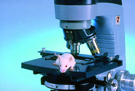
Roughly 60% of the 12m animal experiments in Europe each year involve mice. But despite their undoubted usefulness, mice haven’t been much help in getting successful drugs into patients with brain conditions such as autism, schizophrenia or Alzheimer’s disease. So too have researchers grown 2D human brain cells in a dish. However, human brain tissue comprises many cell types in complex 3D arrangements, necessary for true cell identity and function to emerge.
Researchers are hopeful that lab grown mini-brains – tiny 3D tissues resembling the early human brain – may offer a more promising approach. ‘We first published on them in 2013, but the number of brain organoid papers has since skyrocketed, with 300 just last year,’ says Madeline Lancaster at the Medical Research Council’s Laboratory of Molecular Biology lab in Cambridge. Lancaster was the first to grow mini-brains – or brain organoids – as a postdoc in the lab of Juergen Knoblich at the Institute of Molecular Biotechnology in Vienna, Austria (Nature, doi: 10.1038/nature12517. doi:10.1038/nature12517). The miniature brains comprised parts of the cortex, hippocampus and even retinas, resembling a jumbled up brain of a human foetus.
‘We were stunned by how similar the events in the organoids were to what happens in a human embryo,’ says Knoblich. To be clear, the brain tissue is not a downsized replicate. Lancaster compares the blobs of tissue to an aircraft disassembled and put back together, with the engine, cockpit and wings in the wrong place. ‘The plane wouldn’t fly, but you can study each of those components and learn about them. This is the same with brain organoids. They develop features similar to the human brain,’ she explains.
They used a recipe of growth factors to convert skin cells into induced human pluripotent stem cells, a technique that had won other researchers the Nobel prize in physiology or medicine in 2012. The Vienna lab put these stem cells into a broth made with a nutrient recipe essential for brain development. The stem cells differentiated into embryonic neurons and after a few weeks grew into brain-like tissue 3-4mm across.
The initial skin cells were taken from patients with microcephaly – a neurological disorder resulting in unusually small heads - to create mini-brains, which showed that premature differentiation of neurons underlies this disorder. What was true in the patients, was true in the mini-brains derived from them.
This ability to take cells of patients and convert them into brain tissue with the same genetics is allowing researchers to ask questions difficult to address in mouse models. Knoblich believes epilepsy is partly caused by neurons not migrating correctly in the developing brain of a foetus. He can track the migration of these cells in mini brains derived from epileptic patients, compared with those from healthy people, and discover genes turned on or off only in patients. This could shed light on the mystery of what happens in the foetal brain to lead to epilepsy later in life. In future it may even be possible to repair a mutation in these mini brains to reveal its role in the disorder. ‘The final goal with these mini brains would be to develop models for disorders such as epilepsy, autism and schizophrenia,’ Knoblich explains.
|
300 |
|
In 2014, neuroscientist Rudolph Tanzi at Harvard Medical School in Boston, US, grew human neural stem cells into organoids, which were given two mutations recognised in patients with inherited Alzheimer’s disease. Within weeks, they grew into mini-brains that formed beta-amyloid plaques and tangles of tau proteins – something previously very difficult to achieve with mouse models. |
The potential of mini-brains was quickly recognised by others. ‘Mini brains can be produced from stem cells, including from patients with certain diseases. This is a big step forward,’ explains Thomas Hartung, toxicologist at Johns Hopkins University in Baltimore, Maryland. He has used mini brains to study how viruses such as Zika and HIV replicate inside human brain tissue – something that was not possible in mouse brains. In 2016, Brazilian researchers showed that Zika virus, though not the related Dengue virus, killed human neural stem cells in mini brains, stalled the formation of early brain structures and reduced the overall growth of the mini brains (Science, doi: 10.1126/science.aaf6116).
Mini brains contain different types of brain cells, reflecting conditions in the early human foetal brain. ‘We see all the relevant neurons in these mini-brains, as well as helper cells like astrocytes,’ says Hartung, who has begun using them to investigate possible causes of autism. The Centre for Disease Control (CDC) estimated that one in 59 children had autism in 2014, up from one in 150 in 2002. Hartung believes certain individuals are susceptible while in the womb to known neurotoxic chemicals, such as brominated flame retardants used in furniture or the pesticide chlorpyrifos.
‘Our hypothesis is that it cannot just be chemicals alone,’ says Hartung, pointing out that around 100 genes may contribute to autism. ‘The approach we are pursuing is to produce mini brains that come from autistic children and then look to see whether [the mini brains] have increased susceptibility to certain chemicals.’
He is also mutating certain genes linked to autism to see their effects. Already, he has introduced mutations in a gene involved in early brain development in mini brains derived from a healthy donor. He compared mini brains with either one copy or both copies of this mutated gene and looked at the response to various chemicals. The results have not yet been published, but illustrate the potential of mini brains to study the impact of gene mutations and chemicals in living human brain tissue. ‘Autism is really an epidemic,’ says Hartung. ‘Studying gene-environment interactions in autism has not been possible in any other type of model.’
Even though mini brains are embryonic brain tissue, researchers have begun to use them for age-related conditions such as late-onset Alzheimer’s. This is surprising since mini brains are usually grown for two to three months, whereas Alzheimer’s happens mostly to people over age 65. ‘A few years ago, when we first described brain organoids, I would have said I didn’t think it was possible to study neurodegeneration [using mini brains], since these are a model of the developing brain, not the adult brain,’ says Lancaster.
Nonetheless, researchers were willing to try options other than rodent models for Alzheimer’s and other conditions. ‘Drugs seemed to be working in mouse Alzheimer’s models, and improving things in the mouse brain, but this did not translate into human patients,’ says Vanessa Hall, neuroscientist at the University of Copenhagen, Denmark. Then, in 2014, neuroscientist Rudolph Tanzi at Harvard Medical School in Boston, US, grew human neural stem cells into organoids, which were given two mutations recognised in patients with inherited Alzheimer’s disease (Nature, 2014, 515, 274). They grew into mini-brains that formed beta-amyloid plaques after six weeks. The surprise came around two weeks later: tangles of tau proteins formed in the mini-brains too. This is remarkable because it has always been difficult to get mouse models of Alzheimer’s disease to develop these two disease hallmarks, particularly tau tangles.
Tanzi’s mini brains had two types of tau, like the human brain, whereas mice only ever have one dominant type. Even a layer of human cells cultured in a dish did not create the right mix of tau proteins. ‘Unless you grow human neurons in a 3D culture system, you won’t see the true tau tangles forming,’ Tanzi explains.
In 2016, a study reported that ordinary cells from patients with Alzheimer’s disease could be converted into induced pluripotent stem cells, grown into brain organoids, and shown to produce amyloid plaques and tau tangles (PLOS One, doi: 10.1371/journal.pone.0161969). The researchers went on to show that inhibitors of certain enzymes reduced plaques and tangles, a promising result. ‘These 3D models bring a lot to the table. We use human cells and we can use all kinds of patient-derived cells to create these systems. We can model familial (inherited) and late-onset (sporadic) Alzheimer’s disease,’ says study author Li-Huei Tsai, a neurobiologist at Massachusetts Institute of Technology (MIT) in Boston.
Image: Southern Illinois University / Science Photo Library
There is still more work to be done to improve mini brains for Alzheimer’s. One notable shift in Alzheimer’s research is a greater emphasis on the role of neuroinflammation. ‘If the brain’s innate immune system does not react to tangles and plaques with inflammation, you don’t get dementia.’ says Tanzi. He argues that we need to stop inflammation in the brain to help Alzheimer’s patients, but that the lab mouse will not help us here, since ‘the mouse innate immune system is so different from the human.’
Human mini brains do contain astrocytes, but lack immune cells called microglia. These are made in bone marrow and migrate into the brain. Earlier this year in 2018, Tanzi and colleagues reported how they placed microglia in a reservoir outside a dish with mini brains, and then observed microglia moving towards the mini brains in response to inflammatory signals from stressed neurons (Nature Neuroscience, 2018, 21, 941-51). Hartung agrees that replicating the immune system in mini brains is crucial. ‘Inflammation is really critical, because there is no brain disease that doesn’t have an inflammatory component,’ he explains. ‘We are looking to include microglia in our [mini brain] models of autism, because inflammation is a key factor in this condition.’
Hall in Copenhagen is yet another researcher using mini brains to study Alzheimer’s disease. She is especially interested in cells called stellate neurons, which seem to be impacted very early in the disease. ‘We are interested in making these cell types in a dish, since this is where Alzheimer’s disease always seems to start. We are hoping we can learn more about why it originates there, and about basic mechanisms,’ says Hall. This touches on another strategy to improve mini brains – by altering their growing conditions. Lancaster, for example, is tweaking the recipe of her nutrient broth to nourish neural stem cells to encourage cerebral cortex growth, the area responsible for human cognition and language.
Mini brains are even playing a role in cancer studies. Earlier in 2018, Knoblich developed a mini brain for researching human brain tumours (Nature Methods, 2018, 15, 631). He used Crispr-Cas9 techniques to gene edit mutations into human brain cells to initiate formation of a glioblastoma, a malignant brain tumour with very poor survival rates that have not improved for decades. Senator John McCain in the US was a recent victim. The new mini brain allowed researchers to study tumour biology and growth in a dish, but also test potential drugs against them.
|
Earlier in 2018, researchers reported developing a mini brain for researching human brain tumours. By gene editing mutations into human brain cells to initiate formation of a glioblastoma, a malignant brain tumour with very poor survival rates that have not improved for decades, they were able to study tumour biology and growth in a dish - and screen potential drugs against them. |
Hartung too is using mini brains to study brain tumours, by seeding organoids with tumour cells and observing how they take over. ‘By calibrating the right numbers of cells, we can get to a situation where we can test chemotherapy to see how it can damage the tumour but not the surrounding tissue,’ he explains, citing research not yet published.
Steady progress is being made to commercialise mini brains as a tool for drug discovery. In 2016, Hartung reported the first mass produced and standardised mini brains at the AAAS meeting in Washington DC. ‘This would allow pharma to test the properties of substances for brain diseases, using a standard model,’ says Hartung. A strict protocol generated mini brains around 350 micrometres across, with about 1000 cells. He then launched a company, which faltered for business reasons, but he welcomes the success others have had: a company called Stemonix now sells mini-brains as highly homogenous brain spheres for high throughput drug screening.
New tools are certainly required. ‘There’s been an enormous number of stroke trials that all failed and that can be traced to a failure of animal models to pick the right candidates,’ Hartung says. ‘It makes sense to use humanised models.’ The brain organoids also cost less than lab animals. Yet another advantage is that significant quantities of mini brains can be grown relatively cheaply, opening the door to large-scale screening of potential therapies. ‘Pharmaceutical companies are interested in using these models to test lots of up and coming drug compounds, because these are human and may be better than some of their rodent models,’ says Hall. She noted early on how they could be used to uncover novel mechanisms of Alzheimer’s, which could lead to new drugs, and for testing new compounds (J. Clinical Med., doi: doi:10.3390/jcm3041402).
At Harvard, Tanzi is testing over 1000 US Food and Drug Administration (FDA)-approved drugs in Alzheimer’s disease mini brains to see which ones reduce inflammation and/or tau tangles. So far, he has identified a few dozen that reduce tangles significantly. The future popularity of mini brains seems like a sure bet. ‘What’s really exciting,’ says Hartung, ‘is that the FDA is embracing this technology and they set the standard worldwide. When the FDA says something is good, everyone wants it.’


